
Dens (anatomy): Difference between revisions
mNo edit summary |
m robot Modifying: pt:Processo odontoide |
||
| Line 52: | Line 52: | ||
[[Category:Bones of the torso]] |
[[Category:Bones of the torso]] |
||
[[pt:Processo |
[[pt:Processo odontoide]] |
||
Revision as of 20:46, 3 May 2009
| Dens (anatomy) | |
|---|---|
 Second cervical vertebra, or epistropheus, from above. (Dens labeled at center top.) | |
 Second cervical vertebra, epistropheus, or axis, from the side. (Odontoid process labeled at center top.) | |
| Anatomical terms of bone |
The dens or odontoid process or odontoid peg of the axis exhibits a slight constriction or neck, where it joins the body.
On its anterior surface is an oval or nearly circular facet for articulation with that on the anterior arch of the atlas.
On the back of the neck, and frequently extending on to its lateral surfaces, is a shallow groove for the transverse atlantal ligament which retains the process in position.
The apex is pointed, and gives attachment to the apical odontoid ligament; below the apex the process is somewhat enlarged, and presents on either side a rough impression for the attachment of the alar ligament; these ligaments connect the process to the occipital bone.
The internal structure of the odontoid process is more compact than that of the body.
The odontoid peg is the ascension of the atlas fused to the ascension of the axis. The peg has an articular facet at its front and forms part of a joint with the anterior arch of the atlas. It is a non-weight bearing joint. The alar ligaments, together with the apical ligaments, are attached from the sloping upper edge of the odontoid peg to the margins of the foramen magnum. The inner ligaments limit rotation of the head and are very strong. The weak apical ligament lies in front of the upper longitudinal bone of the cruciform ligament, and joins the apex of the deltoid peg to the anterior margin of the foramen magnum. It is the fibrous remnant of the notochord.
Fracture types
Type I Fracture - Extends through the tip of the dens.
Type II Fracture - Extends through the base of the dens. It is the most commonly encountered fracture for this region of the axis.
Type III Fracture - Extends through the vertebral body of the axis. [1]
Additional images
-
 Articulation between odontoid process and atlas.
 -
 Median sagittal section through the occipital bone and first three cervical vertebræ.
 -
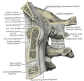
 Sagittal section of nose mouth, pharynx, and larynx.


External links
- Odontoid+process at the U.S. National Library of Medicine Medical Subject Headings (MeSH)* Diagram at umich.edu (Odontoid process is #3)
- Atlas image: back_bone11 at the University of Michigan Health System - "C2, 2nd Cervical Vertebra or Axis - Superior View (Dens is #1)"
- Atlas image: back_bone12 at the University of Michigan Health System - "C2, 2nd Cervical Vertebra or Axis - Posterior View (Dens is #1)"
- Anatomy photo:31:st-0204 at the SUNY Downstate Medical Center - "Pharynx - Bones"
- Walid MS et al: A Variation Of Type III Odontoid Fracture Presenting As Isolated Jaw Pain. The Internet Journal of Orthopedic Surgery. 2007. Volume 7 Number 1. [2]
![]() This article incorporates text in the public domain from the 20th edition of Gray's Anatomy (1918)
This article incorporates text in the public domain from the 20th edition of Gray's Anatomy (1918)
This human musculoskeletal system article is a stub. You can help Wikipedia by expanding it. |